P Bhoopathi, N Lee, A Pradhan, X Shen, S Das, D Sarkar, L Emdad, P Fisher (2016)
mda-7/IL-24 Induces Cell Death in Neuroblastoma
through a Novel Mechanism Involving AIF and ATM
Cancer Research, June 2016 76:12
mda-7/IL-24 Induces Cell Death in Neuroblastoma
through a Novel Mechanism Involving AIF and ATM
Cancer Research, June 2016 76:12
(Translated by Krishna Karamsetty)
Support: Western Blot
|
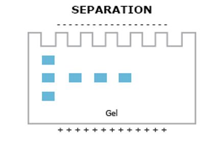
The western blot procedure uses specific antibodies to identify proteins that have been separated by gel electrophoresis and later subjected to a transfer to a specialized membrane where they are analyzed by further more antibodies. In order to this procedure, the researcher collects cells from a culture and kills or lyses them to create a lysate. This lysate or a collection of dead cells is later altered naturally to expose the wanted proteins for analysis. After the lysate has altered naturally or denatured, it is put through a process known as SDS-PAGE which is simply called gel electrophoresis.
|
|
|
Afterwards, the proteins on the gel are transferred to special membrane that is used specifically used for Western Blot through the use of electricity. Later this membrane is blotted with milk to prevent non-specific binding. The milk is used to prevent the primary antibody from binding to the membrane because this could change results and mess up the analysis.
|
|
|
Once the membrane has been covered in several primary antibodies that interact with the wanted proteins in the membrane, in this case, the antibodies that interact with MDA-7 Melanoma differentiation-associated gene-7, the main gene for this project, E1A Gene that allows the virus to replicate and beta-actin. When the researchers are growing the primary antibody, a specific type of animal may be used whether it is a mouse, rabbit or rat. Later on, during the experiment, the general secondary antibody that is used will correspond to the animal that the primary antibody was grown in. The primary antibodies allow the researcher to detect the level of protein on the membrane. After the primary antibodies have thoroughly coated the membrane, the researcher adds a secondary antibody that corresponds to the animal that the primary antibody was grown in. The secondary antibody is also attached with an enzyme known as HRP or horseradish peroxidase. After the attached secondary antibody has thoroughly covered the membrane, it is covered by a chemiluminescent reagent called luminol and hydrogen peroxide which acts as an oxidizing agent. The luminol solution allows the researcher to analyze how much of the protein was created by the lysed cells. This happens because when the luminol solution comes into contact with HRP, HRP catalyzes the reaction between luminol and hydrogen peroxide which leads to a light as the luminol decays.
|

|
|
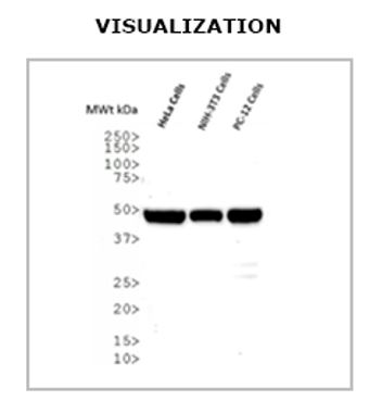
The membrane is then developed in a dark room to get the bands that can later be analyzed. These bands are analyzed by the thickness. The thicker the bands, more expressed that protein is in the original lysate. For this article, the researchers are looking for the bands correlated with MDA-7. When the band is thicker, this means that the virus has successfully injected the MDA-7 gene into the cell and protein production has begun which will cause cell death.
|
|
References
- “Overview of Western Blotting.” Thermo Fisher Scientific, Thermo Fisher Scientific, www.thermofisher.com/us/en/home/life-science/protein-biology/protein-biology-learning-center/protein-biology-resource-library/pierce-protein-methods/overview-western-blotting.html..