P Bhoopathi, N Lee, A Pradhan, X Shen, S Das, D Sarkar, L Emdad, P Fisher (2016)
mda-7/IL-24 Induces Cell Death in Neuroblastoma
through a Novel Mechanism Involving AIF and ATM
Cancer Research, June 2016 76:12
mda-7/IL-24 Induces Cell Death in Neuroblastoma
through a Novel Mechanism Involving AIF and ATM
Cancer Research, June 2016 76:12
(Translated by Krishna Karamsetty)
Experiment: Experiment 3 - Cancer-terminating virus containing MDA-7uses pathways independent of normal cancer killing pathways
|
This experiment was done to demonstrate that the cancer-terminating virus containing MDA-7 was using pathways that are separate of normal cancer killing pathways. Each of one the procedures were done three times in different cells linesCells grown from a singular cell therefore having the same genetic structure which produced the same results. Normal cancer killing pathways involve proteins called caspases or more specifically caspase-3 and caspase-9. Caspases are a group of proteins that cause programmed cell death. When caspases are made, they are inactive, but when the appropriate stimulus arises, they are activated, they start a domino effect which leads to cell death. Caspase-9 initiates the process of cell death while caspase-3 actually carries it out. As soon as caspase-9 was activated, it started a chain reaction activating caspase-3 which can go and start to break down the cell’s essential functions.
|
|
|
|
|
|
For Figure 3A, neuroblastoma cells in petri dishes were infected with either no viruses, the viruses that cannot replicate, the viruses that can replicate but, in all cells, regardless of cancer, and the viruses that can replicate only in cancer cells for 72 hours. After 72 hours, the lysateA collection of dead cells was collected and put through a Western Blot for a different cell death indicator than before using antibodies specific for that indicator. Like Figure 2C if the cell death indicator is cleaved, this means that the cell has or is going through cell death. However, the normal cell death indicators that are involved in programmed cell death aren’t showing up in the Western Blot, but we do know that the virus is successfully doing its job. Since there aren’t two lines for any treatment involving the virus, this made the scientists come to the conclusion that the virus is using a pathway that is separate from the normal cancer killing pathways.
|
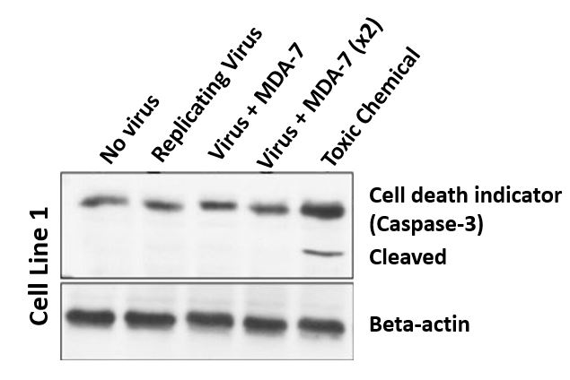
Figure 3A
|
|
|
|
|
For Figure 3B, the cells are infected the same way as 3A for 72 hours. Once the cells have been incubated, they are collected into a lysateA collection of dead cells and put through an assay to measure the level of the cell death indicator. As shown here in the bar graph, the cells that were exposed to the virus have a low level of the classic cell death indicator while the cells that were exposed to the toxic chemical have much higher levels. This further supports the idea that MDA-7 is using a different pathway than normal.
|
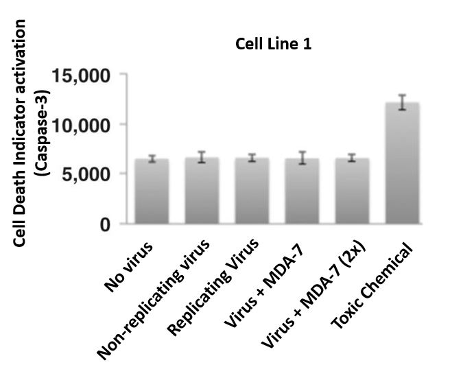
Figure 3B
|
|
|
|
|
For Figure 3C the cells are infected the same way as 3B but they are also treated with a cell death inhibitor as well. Once the cells were incubated with both the treatment and the cell death inhibitor, they were collected into a lysateA collection of dead cells and put through a Western Blot with antibodies specific for the cell death indicator. If the first row of blots has two lines, that means that the cell death indicator that was used in experiment 2 was cleaved but when there is only one line, it means that the cell didn’t go through cell death. This provides further evidence to show that when the inhibitor isn’t present, MDA-7 still starts cell death which further promotes the theory that MDA-7 uses separate pathways than normal.
|
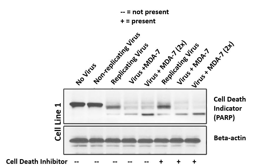
Figure 3C
|