P Bhoopathi, N Lee, A Pradhan, X Shen, S Das, D Sarkar, L Emdad, P Fisher (2016)
mda-7/IL-24 Induces Cell Death in Neuroblastoma
through a Novel Mechanism Involving AIF and ATM
Cancer Research, June 2016 76:12
mda-7/IL-24 Induces Cell Death in Neuroblastoma
through a Novel Mechanism Involving AIF and ATM
Cancer Research, June 2016 76:12
(Translated by Krishna Karamsetty)
Experiment: Experiment 4 - Cancer-terminating virus containing MDA-7 promotes programmed cell death in neuroblastoma
|
This experiment was done to demonstrate that the cancer-terminating virus containing MDA-7 was promoting programmed cell death in neuroblastoma. Each of one the procedures were done three times in different cells linesCells grown from a singular cell therefore having the same genetic structure which produced the same results. In the previous experiment, it was shown that MDA-7 uses pathways that are separate of normal cancer killing pathways. So how exactly is MDA-7 killing these cells. This is where AIF comes in. AIF or apoptosis (programmed cell death) inducing factors is a protein that starts cell death separately from caspases. It does this by fragmenting DNA. AIF also regulates the mitochondriaPowerhouse of the cell. Since the mitochondria is separate from the nucleus of the cell, it wouldn’t make sense for the protein to be found in the nucleus causing cell death. However, when the mitochondria is damaged, AIF moves into the nucleus and starts the process of cell death.
|
|
|
|
|
|
For Figure 4A, neuroblastoma cells in petri dishes were infected with either no viruses, the viruses that cannot replicate, the viruses that can replicate but, in all cells, regardless of cancer, and the viruses that can replicate only in cancer cells for 72 hours. After 72 hours, the lysateA collection of dead cells was collected and put through a Western Blot for cell death inducing factors using antibodies specific for those factors. In the first row, all but 2 of the treatments have two rows of blots. When these cell death inducing factors start cell death, they are cut. When the lysate is put through Western Blot, the antibody binds to both pieces of the factors thus the two lines. When the two lines show up on a Western Blot, it means that the factors are present and working correctly to induce cell death.
|
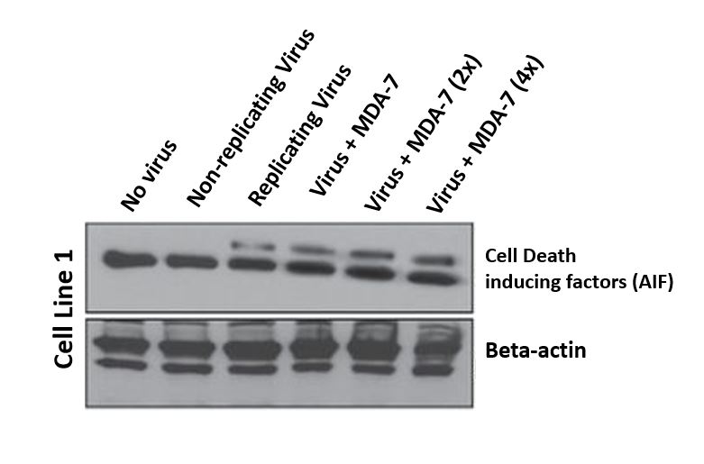
Figure 4A
|
|
|
|
|
For Figure 4B, the cells are infected the same way as 4A for 72 hours. Once the cells have been incubated, they are collected into a lysateA collection of dead cells and put through an immunofluorescence analysis of the cell death inducing factors. Both rows of pictures show that the inducing factors are present regardless of the treatment however when the neuroblastoma cells are exposed to the virus with all the necessary parts, the analysis becomes much clearer and brighter. This increased brightness means that when the cell is undergoing cell death because of MDA-7, these cell death inducing factors are the main pathways that are being used.
|
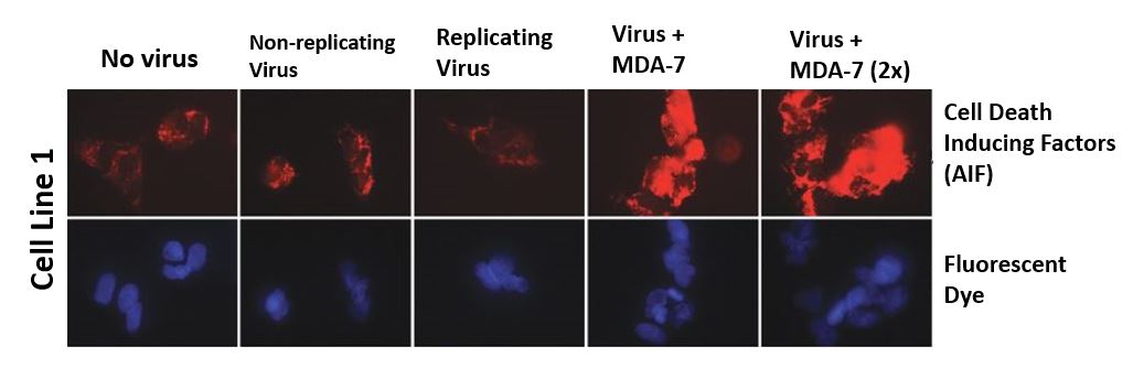
Figure 4B
|
|
|
|
|
For Figure 4C, the cells were only treated with no virus at all, the virus that can replicate in every cell and the virus that can only replicate in cancer cells for 72 hours. Once the incubation period had passed, the cells were collected and put through a Western Blot specifically for the cell death inducing factors. The Western Blot was done for two areas of the cell, the cytoplasm and the nucleus. Every box has four blots in it but only in the nuclear one is the cell death inducing factors more heavily expressed when the cells were exposed to the complete cancer killing virus versus how the incomplete virus and no virus at all had a much weaker expression in the cytoplasm. This means that when the virus had infected the cell with the viral genetic code and MDA-7, the cell death inducing factors are activated and head into the nucleus to begin the process of cell death.
|
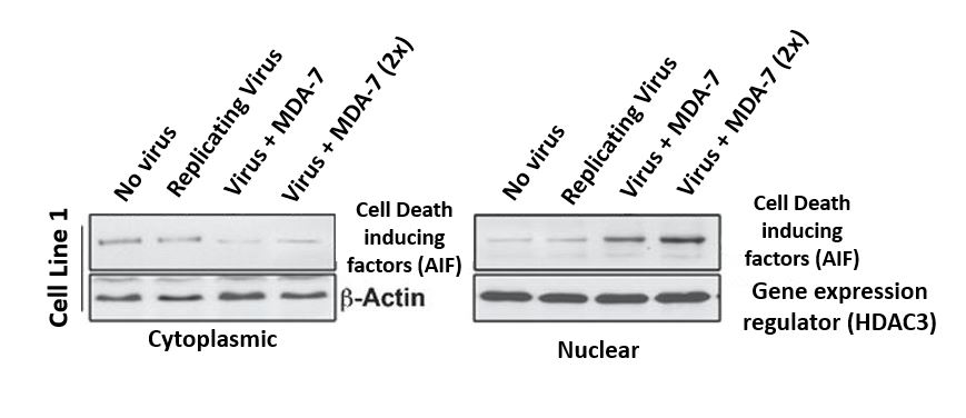
Figure 4C
|
|
|
|
|
For Figure 4D, the cells were first treated with a cell death inducing factors inhibitor and later treated with either no virus, virus that can replicate in any cell and last but not least, virus that can only replicate in cancer cells for 48 hours. After the incubation period, the cells were collected and run through a Western Blot specifically for the cell death inducing factors and the indicator. All three rows of blots show that the factors are present in every case. However, when looking at the inducing factors, when the inhibitor isn’t present, the expression is stronger and even stronger under the virus + MDA-7 treatment and weaker where the inhibitor is present. The cell death indicator is strongly present in all the treatments even when the inhibitor is present however the expression is stronger when the inhibitor isn’t present and when the virus is complete. This shows that the inducing factors play an important role in the strength of the MDA-7 gene when going through cell death.
|
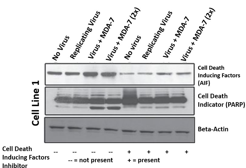
Figure 4D
|