P Bhoopathi, N Lee, A Pradhan, X Shen, S Das, D Sarkar, L Emdad, P Fisher (2016)
mda-7/IL-24 Induces Cell Death in Neuroblastoma
through a Novel Mechanism Involving AIF and ATM
Cancer Research, June 2016 76:12
mda-7/IL-24 Induces Cell Death in Neuroblastoma
through a Novel Mechanism Involving AIF and ATM
Cancer Research, June 2016 76:12
(Translated by Krishna Karamsetty)
Experiment: Figure 6 - Cancer-terminating virus containing MDA-7 works in animals that have grown neuroblastoma tumors
|
Experiment 6 is to show that the virus works inside animals as well. Experiments 1-5 were done in petri dishes or in-vitro, experiment 6 is done inside animals or in-viro. The scientists used a special kind of mouse called a nude mouse. These mice had a compromised immune system where they had a reduced number of T-cells. This was done so that when the human neuroblastoma cells and the virus would receive no resistance from the mouse’s immune system. Two tumors were implanted into the back side of the mouse on both the left and right side of the mouse. However, only the left side was treated. The goal was to see if the virus and MDA-7 after successfully killing the left-side tumor would travel through the blood stream and attack the right-side tumor.
|
|
|
|
|
|
Figure 6A is showing where there was significant tumor reduction in either the left or right side of the mouse. There is significant evidence that the mice treated with the virus+ MDA-7 had a significant tumor decrease versus the mice that weren’t treated at all or the mice that were treated with just the replicating kind. Although tumor on the left-side that were treated with the purely replicating virus did show a decrease, only the tumors that were treated with the virus + MDA-7 showed tumor decreases in the right-side as well. These results confirm a previously established theory that MDA-7 exhibits antitumor “bystander” activity. This means that the gene can travel through the blood stream and induce cell death in tumors away from the original location.
|
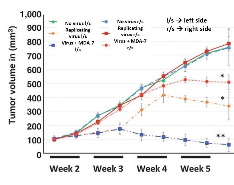
Figure 6A
|
|
|
|
|
Figure 6B shows the same thing as 6A however this time a TUNEL assay was done to show to what extent the cell death was happening. It is evident that when the left side tumor was injected with the virus + MDA-7, cell death is happening very quickly and it also shows that although no treatments were injected into the right side, the virus and MDA-7 travel through the blood stream and start killing the cells there as well. Same with the stain, where the tumor was injected with the virus + MDA-7 there is more cell death as indicated by the increase in purplish, lavender smears throughout the picture. The fluorescent dye also shows that where the tumor was infected with the virus + MDA-7, there is more blackness and less blue and the same shows for the right side as well. This shows evidence that the bystander effect holds true.
|
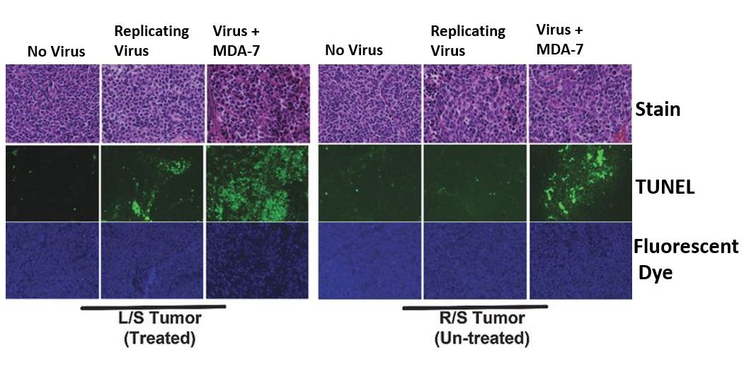
Figure 6B
|
|
|
|
|
Figure 6C shows the same thing as 6A and 6B. After the cells were infected, they were stained with a brown dye. The brown dye interacted with the dying cells giving the images. It is quite prevalent that when the virus + MDA-7 entered into the tumor cells, it rapidly killed the cells which shows that the virus and MDA-7 are successfully doing their jobs. Although the brown color is not as pronounced in the right side as it is in the left, the color is still there, more than the no virus and replicating virus category. Once again this further cements the idea that MDA-7 exhibits a sort off antitumor “bystander” effect.
|
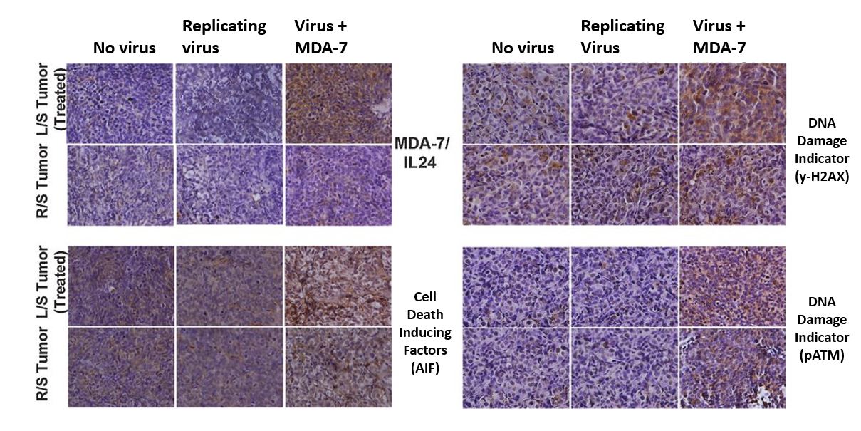
Figure 6C
|