Journal of Comprehensible Results

Hires SA, Zhu Y, Tsien R (2008)
Optical measurement of synaptic glutamate spillover and reuptake
by linker optimized glutamate-sensitive fluorescent reporters
Proc Natl Acad Sci USA 105:4411-4416

(Translated by Jeff Elhai)

Introduction

Nerves carry their messages within the brain and throughout the body through electrical transmission, until the signal reaches the end of a nerve. Then the signal is passed to the next nerve (or muscle) through chemicals called neurotransmitters. Packets of neurotransmitters are released from a nerve ending across a short distance (the synapse) where it is perceived by the cell on the other side (Fig. 1). Neurotransmitters bind to proteins -- receptors -- which in some cases cause electrical transmission to begin anew in the recipient cell.

Glutamate is the most prevalent neurotransmitter of this sort in the brain [1Zhou Y, Danbolt NC (2014). Glutamate as a neurotransmitter in the healthy brain. J Neural Transm 121:817.]. However, glutamate breaks this classical model of neurotransmitter action by spilling out from the synapse and diffusing to nearby cells, where it is detected by glutamate receptors on their membranes. In this way, glutamate can act on a neighborhood of cells, not just the cell across the synapse [2Pal B (2018). Involvement of extrasynaptic glutamate in physiological and pathophysiological changes of neuronal excitability. Cell Molec Life Sci 75:2917-2949.].

To understand how glutamate spillover works, it would be helpful to know how much glutamate is present and at what times during and after nervous stimulation. Several previous attempts have been made to measure glutamate near nerve cells [3-9], however each attempt was deficient in localizing glutamate either in time or space.


Fig. 1: Synapse with neurotransmitter released from synaptic vesicles: Upon stimulation, neurotransmitter is released into the synapse and binds to receptors on the opposing membrane. Some neurotransmitter may spill out of the synaptic cleft and diffuse to neighboring cells. (Image modified from Neurotransmitter (Wikipedia) by Thomas Splettstoesser [CC BY-SA 4.0])
 
Recent technological advances have made possible more accurate measurements of glutamate in and near cells [10Tsien RY (2005) Building and breeding molecules to spy on cells and tumors. FEBS Lett 927–932.,11Okumoto S, Looger LL, Micheva KD, Reimer RJ, Smith SJ, Frommer WB (2005) Detection of glutamate release from neurons by genetically encoded surface-displayed FRET nanosensors. Proc Natl Acad Sci USA 102:8740–8745.]. The method [
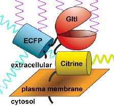
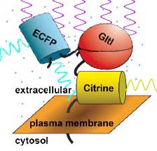
12Deuschle K, Okumoto S, Fehr M, Looger LL, Kozhukh L, Frommer WB (2005). Construction and optimization of a family of genetically encoded metabolite sensors by semirational protein engineering. Prot Sci, 14:2304–2314.], illustrated in Fig. 2 relies on two proteins that are intrinsically fluorescent, fused to a third protein that controls the proximity of the two fluorescent proteins, depending on the presence of glutamate. One protein, Enhanced Cyan Fluorescent Protein (ECFP) fluoresces blue-green light when exposed to violet light. The second, Citrine, a type of Yellow Fluorescent Protein (YFP) fluoresces yellow-green light when exposed to blue-green light. The two proteins were fused to a third -- glutamate periplasmic binding protein (GltI), which changes its shape when it binds glutamate. The concentration of glutamate can therefore be detected by the amount of yellow fluorescence emitted in response to violet light: the more glutamate is present, the more is bound to GltIglutamate-binding protein, and the resulting changein protein conformation pushes ECFPenhanced cyan fluorescent protein away from Citrinea type of yellow fluorescent protein. This leads to less aborbance of blue-green light by Citrinea type of yellow fluorescent protein and less yellow-green fluorescence.

Unfortunately, existing fluorescent glutamate detectors show only a small difference in yellow-green fluorescence in response to glutamate. The authors sought alterations in the structure of the original detector that showed a greater difference and then used this modified detector to monitor glutamate levels near neurons in real time.


Fig. 2: Principle of monitoring presence of glutamate through fluorescent energy transfer:
(LEFT) In the absence of glutamate, violet light excites ECFPenhanced cyan fluorescent protein, which emits cyan light. Because ECFPenhanced cyan fluorescent protein is close to Citrinea type of yellow fluorescent protein, much of the cyan light is absorbed by Citrinea type of yellow fluorescent protein and re-emitted as yellow light.
(RIGHT) In the presence of glutamate, GltIglutamate-binding protein binds to glutamate, altering the position of one fluorescent protein relative to the other. Now when ECFPenhanced cyan fluorescent protein absorbs violet light and emits cyan light, much less of it is absorbed by Citrinea type of yellow fluorescent protein and there is much less emission of yellow light.

References

  1. Zhou Y, Danbolt NC (2014) Glutamate as a neurotransmitter in the healthy brain. J Neural Transm 121:817.
  2. Pal B (2018). Involvement of extrasynaptic glutamate in physiological and pathophysiological changes of neuronal excitability. Cell Molec Life Sci 75:2917-2949.
  3. Clements JD, Lester RA, Tong G, Jahr CE, Westbrook GL (1992) The time course of glutamate in the synaptic cleft. Science 258:1498-1501.
  4. Pasti L, Zonta M, Pozzan T, Vicini S, Carmignoto G (2001) Cytosolic calcium oscillations in astrocytes may regulate exocytotic release of glutamate. J Neurosci 21:477-484.
  5. Copenhagen DR, Jahr CE (1989) Release of endogenous excitatory amino acids from turtle photoreceptors. Nature 341:536-539.
  6. Allen TG (1997) The "sniffer-patch" technique for detection of neurotransmitter release. Trends Neurosci 20:192-197.
  7. Diamond JS, Bergles DE, Jahr CE (1998) Glutamate release monitored with astrocyte transporter currents during LTP. Neuron 21:425-433.
  8. Pomerleau F, Day BK, Huettl P, Burmeister JJ, Gerhardt GA (2003) Real time in vivo measures of L-glutamate in the rat central nervous system using ceramic-based multisite microelectrode arrays. Ann N Y Acad Sci 1003:454-457.
  9. Innocenti B, Parpura V, Haydon PG (2000) Imaging extracellular waves of glutamate during calcium signaling in cultured astrocytes. J Neurosci 20:1800-1808.
  10. Tsien RY (2005) Building and breeding molecules to spy on cells and tumors. FEBS Lett 927-932.
  11. Okumoto S, Looger LL, Micheva KD, Reimer RJ, Smith SJ, Frommer WB (2005) Detection of glutamate release from neurons by genetically encoded surface-displayed FRET nanosensors. Proc Natl Acad Sci USA 102:8740-8745.
  12. Deuschle K, Okumoto S, Fehr M, Looger LL, Kozhukh L, Frommer WB (2005). Construction and optimization of a family of genetically encoded metabolite sensors by semirational protein engineering. Prot Sci, 14:2304–2314.