Journal of Comprehensible Results

Joy Y. Feng, Allison A. Johnson, Kenneth A. Johnson and Karen S. Anderson(2001)
Insights into the Molecular Mechanism of Mitochondrial Toxicity by AIDS Drugs*
The Journal of Biological Chemistry Vol.276,No.26, Issue of June, pp.23832-23837

(Translated by Owais A. Shahzada)

Introduction

The various forms of nucleoside analogsDrugs that inhibit viral replication that have been observed have caused dysfunctions within in the mitochondrial DNA. Some of the dysfunctions that have been observed in the patients are neuropathydysfunction of the nerves causing numbness or weakness, myopathyA disease or disorder that weakens the skeletal muscle, and pancreatitisDisease in which the pancreas is inflamed[2]. These dysfunctions have come from other nucleoside analogs; for instance, AZT, ddI, and ddC. All three are nucleoside analogs causing some form of dysfunction. All the symptoms that have come about seem to be related to the inhibition of DNA polymerase within the mitochondria. The toxic drugs which are taken by the patients have shown to be causing mitochondrial dysfunctions which can be related to mitochondrial genetic disorders, like the ones mentioned above[3]. Being exposed to the nucleoside analogs are affecting the mitochondria by decreasing its ability to create new strands of DNA, the ability for the cell to survive and live successfully, and changes in the overall structure of the cell[4].

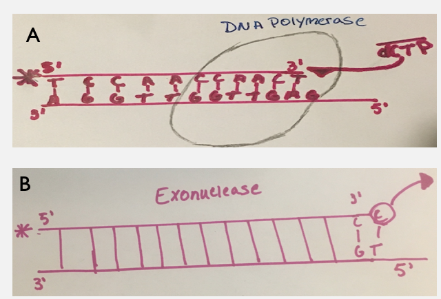
Studies in the past have been done and found that the specific enzyme that is being the most affected is, DNA polymerase gamma[5]. This specific enzyme has two functions, one synthesizes new strands of DNA (polymerase) in 5'-3' direction. The second function is the ability to proofread from the 3' to 5' direction looking for any incorrect mismatches (exonuclease) and replaces it with a correct nucleotide.[6] Past studies that have been performed say that the (-)3TC is a less toxic analog compared to its counterpart (+)3TC[7]. These studies however are not the best because when running the experiments, the researchers used such a small concentration of mitochondrial DNA polymerase that it would not exist as a holoenzyme. With the availability of constructing a pure human mitochondrial DNA polymerase this allows for a deeper and thorough analysis[9].

The main goal of the researchers is to look at the incorporation of an exonucleaseThe polymerase activity which cuts incorrect nucleotides activity, efficiency, and time it takes for the binding to occur. Also, the activity of the nucleoside analogsDrugs that inhibit viral replication is checked to see if it is cutting out the wrong incorporated nucleotide, and the rate at which it is cleaving and adding an analog. Both the enantiomers are compared back to the wild-type known as dCTP (dicytidine triphosphate). Comparing the nucleoside analogs back to the wild-type (dCTP) will help understand how the analogs are incorporating with mitochondrial DNA polymerase and how the polymerase is being inhibited. With the knowledge gained from the experiment this may help to design better antiviral drugs which inhibit the viral DNA polymerase rather than the human mitochondrial DNA polymerase.


Fig. 3: DNA/DNA Nucleotide Sequence: The sequence is are known as oligonucleotides which are short sequences of nucleic acid polymers. The DNA primer/DNA template is a duplex sequence that contains a radioactive phosphate (denoted by an asterisk at the 5 prime position of the DNA for the 23mer sequence) on the primer for visualization of that DNA strand. The analogs in Fig. 2 will bind to the DNA polymerase and be incorporated into the duplex sequence, resulting in an increase in length by 1 nucleotide into the sequence. The phosphorylated sequence 23mer sequence will help identify when the new nucleotide binds into the sequence because it will become 24 nucleotides in length and can be separated on a gel.


Fig. 4: Illustration of DNA polymerase and exonuclease activity: Two images are shown in this figure, Image A and Image B. Image A illustrates a DNA duplex sequence that contains a sketch of a blob. The blob is representing DNA polymerase where the synthesis of DNA is occurring by the incorporation of the wild-type dCTP. Image B represents the exonuclease activity occurring. C does not bind with T, so the C will be cleaved off.

References

  1. Mark Cichocki, RN Updated November 26, 2018. .
  2. Simon D.K.,Johns D.R. Mitochondrial(1999) Disorders: Clinical and Genetic Features. Vol. 50:111-127 (Volume publication date February 1999) .
  3. Lewis W., Dalakas M. C.(1995) Nature Medicine volume 1, pages 417–422 (1995) .
  4. Medina D. J., Tsai C. H., Hsiung G. D., Cheng Y. C.(1994) DOI: 10.1128/AAC.38.8.1824 .
  5. Chang C. N., Skalski V., Zhou J. H., Cheng Y. C.(1992) Biochemical pharmacology of (+)- and (-)-2',3'-dideoxy-3'-thiacytidine as anti-hepatitis B virus agents. The Journal of Biological Chemistry 267, 22414-22420.
  6. Kukhanova M., Liu S.-H., Mozzherin D., Lin T.-S., Chu C. K., Cheng Y.-C.(1995) J. Biol. Chem. 270:23055–23059..
  7. Graves S. W., Johnson A. A., Johnson K. A.(1998) Biochemistry, 1998, 37 (17), pp 6050–6058 .
  8. Martin J. L., Brown C. E., Matthews-Davis N., Reardon J. E.(1994) Antimicrob. Agents Chemother. 38:2743–2749. .
  9. Gray N. M., Marr C. L. P., Penn C. R., Cameron J. M., Bethell R. C.(1995) Volume 50, Issue 7, 28 September 1995, Pages 1043-1051.
  10. Gel electrophoresis Khan academy.
  11. KinTek Instruments Model RQF-3 kintekcorp.com.
  12. Densitometer silkscientific.com.