The main purpose of real-time PCR is to amplify the regions of a gene that one may want to analyze through quantification. In this case, amplification of sections of the DNA in real-time forward and reverse for p21, p53, PDGFR (alpha and beta). This is done through the utilization of an enzyme that can go through a single strand and undergo base synthesization. Primers are utilized to initiate where in the sequence the amplification should begin. The polymerase enzyme then binds and replicates the sequence onwards. Afterwards, two sequences in total are now possessed. These are the principles of normal PCR, within real-time PCR, the samples are observed and recorded through a machine usually with a camera. PDGF and FGF were also treated with sunitinib and SU5402, the two antagonists to diminish the effects of the two factors.
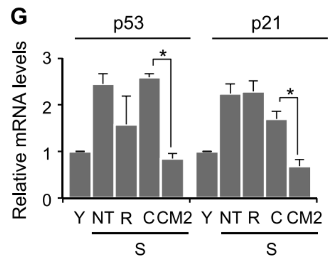
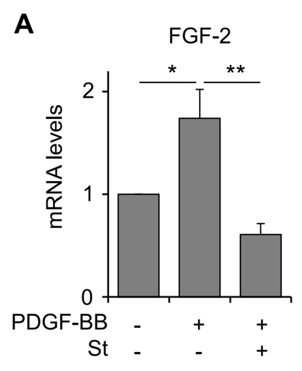
After the process of real-time PCR, the following image was then presented on the right showing mRNA levels. A reduction in protein expression was seen in both p21 as well as p53 in mRNA as well as protein expression (Fig. 2G). Their results also demonstrated that PDGF -BB along with treatment with sunitinib decreased mRNA levels of FGF -2 significantly (Fig. 5A). With only PDGF -BB levels increased. This also demonstrates part of their methods showing that sunitinib is doing its job as an antagonist (Fig 5A).
Real-Time PCR Analysis; The Procedure
1. Bae et al. first took their ESCs and extracted the RNA utilizing a Tri-RNA isolation reagent. This reagent is from Life Technologies. The way this isolating reagent works on the stem cells is through breaking down the cell as well as homogenizing it, in a manner so that all that remains is solely the RNA in an aqueous state. This aqueous state isnt just a result of adding this reagent but occurs after centrifugation. They perform centrifugation to further isolate the RNA resulting in this aqueous state. However, there is still organic material that may sometimes be seen within the microfuge tube, so chloroform is usually added to break down further organic material resulting in RNA being the only substance within the tube.
2. With the RNA isolated from the ESCs, they can now move onward to amplification of cDNA with their gene-specific primers to amplify specifically PDGF receptor alpha and beta as well as FGF. cDNA is generated from the isolated RNA through the utilization of reverse transcriptase, which synthesizes one cDNA strand, the complement to that strand is usually produced through enzyme RNase H. They then had to melt the DNA into separate strands for the primers to bind onto, this is usually done by raising the temperature to about 95 degrees Celsius. After the DNA strands are separated, the temperature is then lowered to about -60 degrees Celsius in which then the primers are added to the cDNA strands. The primers amplify the region of cDNA through moving from the 5 prime to 3 prime on each end of the DNA strands towards the center of the region. The primers dont just bind anywhere, but rather where their corresponding specific gene of interest is located. For this experiment, it is not mentioned how many primers are utilized. After this, a polymerase enzyme will come to synthesize that strand onwards. This is usually done around 70 degrees Celsius to accelerate the enzyme. This cycle is done usually around 40 times, yielding billions of DNA strands.
3. With Real-time PCR, the same steps are performed, but the DNA is rather stained with a fluorescent dye so that the process of amplification can be monitored by a specific camera within a machine.
4. The main benefits of real-time PCR that it gives you a live look at the reaction so you can tell which genes are being expressed and which ones are not, in this case, genes for p53, p21, PDGF, and FGF. This was part of the reason they performed a real-time PCR along with the fact that PCR real-time data can provide a truly quantitative model due to the ability to look at gene expression in real time. The specific lab machine they utilized in this scenario was a 7500 Real-Time PCR system. Usage of this 7500 system usually generates results within 30 minutes.
5. However, they amplified the genes for p53 and p21 slightly different from PDGFR and FGF. They utilized SYBR Green, which is an intercalating dye. Intercalating meaning an insertion dye. SYBR Green is a dye that binds to DNA helix as a whole. Thus, by altering the structure, this increases fluorescence within the DNA sample. As to why this was used for p53 and p21 but not the others, it is not mentioned. However, it could be possible that the fluorescence is harder to observe within p53 and p21 so they have to add SYBR Green.
6. I have included further support figures within the support section for Real-Time PCR.