
The following are interesting images, photos, and figures from neurogenetics research. I initially had more images posted here, but took a few off for ethical, copyright, and confidentially reasons. Currently, the only brain images are my own, though I'll soon add a picture of my wife's brain activation during a fMRI music task (once the data is published). Hopefully as I continue research at VIPBG, I'll have some more interesting things to post here.
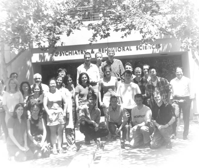 | An old-style picture of SPNL and associated researchers taken July of 2001. The photographer was Ben Krasnow, using an 19th century camera. |
| These images are taken from a high-resolution T1-weighted volumetric (SPGR) MRI scan on a GE 1.5 Tesla scanner, with a slice thickness of 1 mm. It is difficult to tell from coronal sections without practice, but this brain isn't all that remarkable; it's a little smaller than average, but well within typical range. The image resolution has been substantially worsened to make the file of a reasonable size, and to purposefully prevent its use as data. | |
| A surface rendering of my brain using BrainImage's ray tracing function, based on the coronal images seen above. Yes,some may be surprised to find that there actually is something within my skull, but here's the proof. This is one of my favorite images because one really can visualize the overall shape and architecture of the brain, and my gyrification pattern is nearly textbook if I do say so. It's still hard for me to believe that so clear a picture of the brain can be obtained without invasive procedures. This subject is (presumably) still alive and getting along in life, though I sometimes wonder. | |