Anatomy of the Skeletal System
Bone Anatomy
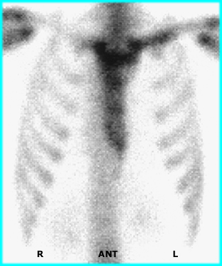
Compare the nuclear medicine scans to anatomical diagrams. What can you label/identify on the NMT exam. Spot views were taken of the chest, spine, hand, and foot.
Chest Anatomy |
Chest Spot View |
 |
 |
Comments on how does one improve the quality of a spot view (Chest) on a bone scan?
- Wait 3 hours before you start the scan
- Have the patient drink lots of water
- Make sure the detector is as close to the patient as possible. Increased distance, increases scatter, and reduces resolution
- Use a 256 x 256 matrix. You've got lots of counts so use a large matrix size
- High count imaged require the use of a HR collimator
Anatomy of the Spine - Right Lateral View |
Posterior Spine Spot View |
 |
 |
Comments of spot view of the spine
- The right lateral image defines the anatomy, however, only a posterior is available from an imaging standpoint
- Can the anatomy of the different vertebrae be identified?
- Define the anatomy on the acquired image
Anatomy of the Left Hand |
Three Phase (and Pinhole) Bone Scan of Hand |
 |
 |
Comments of the Three Phase of the hands
- Increased activity is noted in both wrist in all phases of the procedure is an indication of of disease
- However, the anatomy difference from a child to an adult. For this reason the growth plates have increased activity
- Disease is specifically noted as a slight increase in activity in the second metacarpal
- This is an example of a benign bone lesion known as osteoid osteoma
- For more information visit http://www.med.harvard.edu/JPNM/CH/JS2/WriteUp.html
Anatomy of the Foot |
Three Phase Bone Scan of the Feet |
 |
 |
Comment on Three Phase Bone scan of the feet
- Classic for osteomyelitis showing increased uptake in all three phases of the procedure
- This patient also has diabetes mellitus that reduces blood circulation over time (~15-20 years) to the extremities, in turn this will increases the likelihood of infection, followed by amputation
- Is it possible to identify the structures in the foot? Or is there too much background?
- Why is there more background in this procedure as compared to all the others (above)?
- In this specific patient, what might you do to improve target to background?
- For more information about this procedure visit http://www.med.harvard.edu/JPNM/TF99_00/Sept21/WriteUp.html

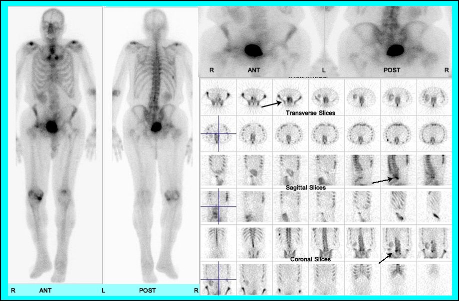
http://ausnucmed.wordpress.com/2009/06/28/pelvic-kidney-on-bone-scan/
- Degenerative changs in the right hip can be noted in the whole body bone scan
- Why is there lack of activity in the left hip?
- However, SPECT imaging also defines increased activity in L5 not seen in whole body and spot views
Return to the beginning of the Document
Return to the Table of Content
Continue to the Next Lecture
8/19