Thyroid - Uptake and Scan

The physiology behind the thyroid is the making of thyroid hormones
  1. Iodine ion (I-) is trapped through the aerobic process via active transport
    1. Trapped iodine concentrations are between 25 (normal) to as high as 5000 (hyper) when compared to the surrounding tissue
    2. The follicular cells in the thyroid organify the iodine by peroxidases in the presence of hydrogen peroxide, converting I- to I2
    3. Tyrosyl and I2 are on the surface of thyroglobulin (TBG) and forms monoiodotyrosine globulin (MIT)
    4. The following hormones are produced within the follicular spaces:
      1. MIT + MIT = Diiodotyrosine. (DIT)
      2. MIT + DIT = Triiodothyronine (T3)
      3. DIT + DIT = Tetraiodothyronine - Thyroxine (T4)
      4. T3 and T4 are bound to TBG and released into the vascular pool as needed
    5. A negative biofeedback loop then regulates production of T3 and T4. As T3 and T4 are generated, TSH and TRH will respond in the opposite manner - known as a negative biofeedback
    6. Radiopharmaceuticals available for imaging
      1. 123I - uptake and scan
      2. 131I - therapy
      3. 99mTcO4- (pertechnetate) can also be used to image the thyroid. While it is not iodine, it does a molecular weight and electronic configuration
  2. Diseases
    1. Hyperthyroidism
      1. Hyperthyroidism results from excess levels of T3 and T4 which can be caused by several factors
      2. Classically, the patient is presented as nervous, fatigued, may show weight loss, proximal muscle wasting, exophthalmos, intolerance to heat, and pretibial myxedema
      3. The cause can be from hyperfunctioning single nodular (Plummer's) within the gland or a multi-nodular goiter usually referred to as Grave's disease - There are several other issues that may cause hyperthyroidism (covered next summer)
      4. To determine hyperthyroidism an iodine uptake procedure may be considered, which calculates the amount of radioactive iodine picked up by the gland
      5. The Calculation for uptake is seen below
    2. Hypothyroidism
      1. Patient will appear fatigued, have cramps, and be constipated
      2. Myocardial contractility is greatly reduced, resulting in a reduction of pulse rate and stroke volume
      3. In severe cases, the patient may be in a comatose state
      4. See uptake and calculation used to determine the disease state
    3. Nuclear medicine has three methods of examining the thyroid
      1. Iodine uptake
      2. Thyroid Imaging
      3. Pharmacological stimulation (cover next summer)
    4. When administering a radiopharmaceutical radiation dosimetry should be considered
      1. A 200 μCi dose of 123I will deliver 2.6 rads to the thyroid and 6 mrads whole-body
      2. A 50 μCi of 131I will deliver 65 rads to the thyroid and 23 mrads whole-body
        1. Note -The administered dose of 131I dose so 1/4 the 123I dose
        2. If a 200 μCi was administered to the patient, why is the radiation burden so different between the two iodines? Answer
      3. A 10 mCi dose of 99mTcO4-will deliver 0.6 rad to the thyroid and 70 mrad whole-body

Radiopharmaceutical used for thyroid uptake and/or scan

Radiopharmaceutical

123I

131I

99mTcO4-

Physiology

Trapped/organified

Trapped/organified

Trapped

Gamma/beta

Gamma

Gamma/beta

Gamma

Dosimetry

Low

High

Low

T1/2

13 hours

8 days

6.02 hours

KeV

159

364

140

Uptake/image

Both

Both

Image only

Therapy

No

Yes

No


  1. Thyroid uptake - Comments on Thyroid Neck Phantom and Positioning
    1. There are two basic formulas/procedures to consider when determining thyroid uptake
      1. The formula below is what you must know this semester
        1. Count thyroid pill in thyroid phantom using uptake probe (distance 20 cm) = Pill count at 0 hr (phantom simulates neck attenuation)
        2. Count Room at 0 hour = Bkg1
        3. Administer the dose patient
        4. Count the patient's thyroid at 20 cm at twenty-four hours = Patient thyroid at 24 hrs
        5. Room background at 24 hours = Bkg2
        6. Apply formula (see below)

thyuptakesimplecorrected2.gif - 9268 Bytes

      1. Example of 123I thyroid uptake calculation:
        1. Pill count at 0 hr = 1,545,667 cpm
        2. Bkg1 = 132 cpm
        3. Patient's thyroid count at twenty-four hours = 215,558 cpm
        4. Bkg2 = 2,154 cpm
        5. Answer = 49.6% or 50%
        6. Decay factor = 0.278
        7. 1,545,667 - 132 = 1,545535 (net counts a 0hr)
        8. 215,558 - 2154 = 213,404 (net thyroid counts at 24 hr)
        9. 1,545,535 x 0.278 = 429,659 (pill count adjusted for 24 hr decay)
        10. (213,404/429,659) x 100 = 49.6 % uptake
  1. Thyroid Scan (for more detail, see Thyroid Procedure):
    1. Matrix size for 123I or 131I = 128 x 128
    2. Matrix size for 99mTcO4- = 256 x 256
    3. Pinhole collimator
    4. Views: ANT; ANT with a marker; RAO; and LAO
    5. 25 to 50k counts with iodine
    6. 100 to 200k counts with 99mTcO4-
    7. ANT marker view - mark the sternal notch and all thyroid nodules
    8. The physician should palpate the thyroid and mark each nodule with a black marker so that the technologist can label it with a radioactive source
    9. Marking the nodule
      1. Take the 57Co flexible marker, circle all the palpable/marked nodules and take the ANT image. This will assist in determining where the whether the nodule is extrinsic or intrinsic to the thyroid
      2. Some clinics only mark the sternal notch, which usually occurs if the physician doesn't palpate the thyroid
  1. Thyroid Imaging (several examples)

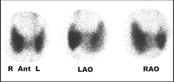
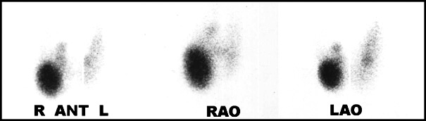
      This is an example of a normal thyroid scan. Three views are taken ANT, RAO, and LAO. No marker image is seen, however, suggested. The marker should be placed at the sternal notch and on any/all nodules noted from thyroid palpation

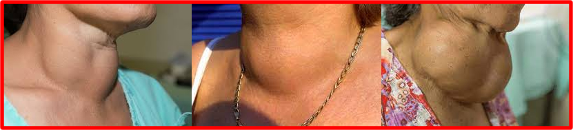
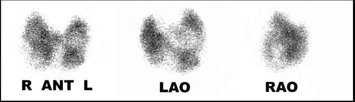
      Before we look at disease from an imaging standpoint, what does a thyroid goiter look like?


      >

      thyroidnormtc2.jpg - 50346 Bytes

  2. The second case is an example of an 123I thyroid scan. The disease that is present is known as Plummer's disease, in which there is a single hot autonomous nodule. This nodule is located in the lower right lobe. Note how the hot nodule has all but shut down normal thyroid function. Why do you think this has happened? Hint - Look at the biofeedback loop

The third case is a thyroid scan that appears large and has heterogeneous uptake. This is an example of multi-nodular disease, in which there are numerous cold and warm nodules

thyroidmutlinodg2.jpg - 34633 Bytes

Thyroid Uptake Procedure
Thyroid Scan with 123I
Return to the Beginning of the Document
Return to the Table of Contents

6/21