 1
1
- PSA density is defined as the amount of PSA per unit volume of prostate tissue. This value is measured by determining the mass of the prostate via a transrectal ultrasound (also known as TRUS). This is usually suggested with patients that have a suspicion for disease, however, TRUS is also suggested when the PSA value is either normal of just slightly elevate. TRUS is determined by comparing the PSA level to the amount of tissue measured. If a patient has a PSA between 4 and 10 ngm/ml and the PSA density is greater than 0.15, there is a 95% sensitivity and a 24% specificity of disease
- PSA velocity is another method to evaluate disease, which is done by evaluating the PSA level over time. If the PSA level increases by greater than 0.75 ng/ml/year then this is considered a positive predictor for developing prostate cancer. If the PSA velocity is 0.15 ng/ml or less per year then this is usually associated with BPH. The suggested way to determine the difference between BPH and cancer of the prostate is to take three samples of blood serum over an 18-month period of time. Also age plays an important factor where increased levels of PSA may actually be associated with BPH
- Testing other types of PSAs (free, bound, Complex, and -[2]Pro) - while there seems to be some improvement in detecting disease when certain forms of PSA are measured, the end result seems to be that specificity does not improve that much
- Age specific ranges for PSA for "normal"
- 40 - 49 years = 0 to 2.5 ng/mL
- 50 to 59 years = 0 to 3.5 ng/mL
- 60 to 69 years = 0 to 4.5 ng/mL
- 70 to 79 years = 0 to 6.5 ng/mL
- This is important, in order to determine the appropriate form of treatment. Perhaps the biggest concern is that 50% of patients, who undergo radical prostatectomy, have tumor that has spread beyond the prostate. Likewise, even with the radical approach for treatment there is a significant incidence of recurrent of disease
- CT, MRI, and TRUS are used to determine if disease is present and whether or not it has spread. However, CT and MRI have a poor predicative value and there are several reasons for this. The issue relates to anatomical imagine. As an example, if if CT/MRI finds an enlarge lymph node or nodes, what is it due to? Inflammation or metastatic disease?
- Regarding TRUS there is improvement in accuracy at the locoregional level, but it serve no use when disease goes distal
- Gleason Grading is used when there is known prostate cancer, which may have been determined by biopsy. Look for term Gleason Grading when referring to the patient's chart. It is an attempt to determine the level of severity of disease
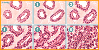
- The Gleason system is based on the architectural patter of tumor cells, specifically prostate carcinoma. The ability for the tumor cells to mimic normal cells is known as differentiation. If a tumor cell is well differentiated, means that it closely resembles the prostate cell, and would receive a Gleason grade of 1 (not a lot of change, therefore the disease isn’t that “bad”). As the tumor cell becomes less differentiated (more abnormal looking), the Gleason scale increases. The highest level or value is 5. Also, the higher the grade, the poorer the prognosis for the patient, and the greater extent of disease
- There is also another term associated with the Gleason grade and it’s called the Gleason score or Gleason sum. Looking for the two most common architectural patterns within the biopsy derives this value. Another words, usually there is more than one pattern of cell. Hence, the pathologist looks for the two most common cell types. If the only type of cell found is a grade one, then 1 and 1 are added together, which gives the Gleason sum of 2. If however, the patterns of cells were mostly of grade 2 and 4, then the patient would receive a score of 6. The highest score that a patient could receive would be a 10, which would be the addition of two grades of 5
- As a general rule a Gleason grade and score assist the clinician on a patient's prognosis , meaning that lower the score the better the prognosis. However, the literature does note that low Gleason scores does not always mean that the patient will do well, likewise there are situations where the patients response well to treatment with no recurrence of metastatic disease, yet the patient had a high Gleason score
- Staging prostate cancer2
- T1 - Tumor is contained with the prostate bed. Not felt by DRE
- T2 - Still confined to the prostate bed, however, it can now be detected by DRE
- T3 - Tumor has spread to adjacent tissues and may include the bladder and seminal vesicles
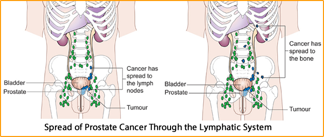
- T4 - Further spread of the tumor to include the lymph nodes and pelvic bone
- See how it spreads
- It starts locally, within the prostate; from there it usually spreads to the surrounding tissues and then lymphatically to the nodes located in the pelvis, and then finally to the bone
- Each stage or progression of disease indicates a poorer prognosis
 2
2

- The treatment of prostate cancer - The problem with treating prostate adenocarcinoma on how soon its detected
- As an example, surgery, via radical prostatectomy done with a localized tumor has a 90% or better five-year survival rate
- However, if the tumor is found extracapsular or it moves beyond the prostate, the ten-year survival decreases to between 40 and 60%
- Also a patient with a Gleason score of 7 or greater has the highest degree of recurrence of tumor
- The main complications for a radical proctectomy are impotence and incontinence with rates being 30 to 90% and 0.5 to 11%, respectively. A surgical technique called nerve-sparing prostatectomy reduces the level of impotence and incontinence
- Forms of therapy include: radiation therapy, brachytherapy, cryotherapy, and delayed androgen deprivation.
- Radiation therapy on patients that have encased tumor have a 80 to 90% five year survival rate, however this number drops to between 65 to 80% at ten years. Complications that occur with this technique, about 5% of cases, the patient may end up with cystitis, urethral strictures, and/or incontinence. In addition there is up to a 15% possibility of erectile dysfunction.
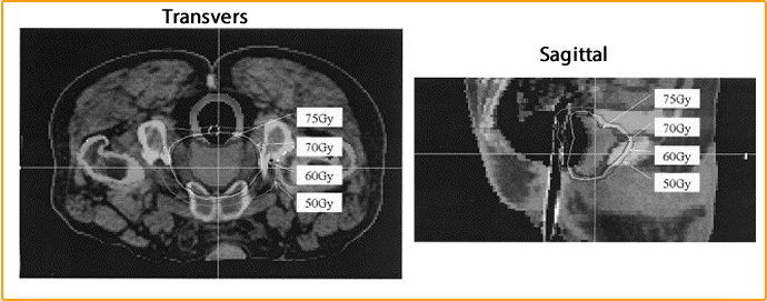
- Intensity-modulated radiotherapy (IMRT) - External beam application shows the distribution of total dose to the prostate and surrounding area. Application of radiation therapy with a Gleason score of less than 7, and a PSA of less than 20 ng/ml will have cancer reoccurrence rate at 4%. While a Gleason score of greater than 7 and a PSA greater than 20 ng/ml will only be 27% cancer free after 3 years
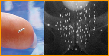
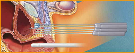
- Seed implants have been around since the early 1900, and currently 125I and 103Palladium are used. Apparently the results of treatment are very similar external radiation beam therapy , however, the complications from treatment seem to be more severe. These severities include: obstructive voiding s symptoms at 27% and perineal pain at 19%. Also few patients may experience urethral necrosis, strictures, bladder constricture and protections. Impotency occurs 10 to 30 % of the time. Seed implants and IMRT can be used in combination
- Cryosurgery is also used in the treatment for localized tumor. This technique has been refined with the use of ultrasound to identify the prostate for guiding the cryoblation procedure. Complications are less than that of radical prostatectomy yet perineal pain, incontinence, and impotence do occur. Data on survival rates cannot be determined at this time because this is a relatively new approach in the treatment of prostate cancer
- Delayed androgen deprivation is the final treatment under this discussion. This procedure is more widely accepted in Scandinavia , in which the testicles are removed. Patients with prostate cancer that has cancer spread beyond the its bed have improved survival rates if they have undergone this therapy. The key being a loss of producing male hormones. Delayed androgen oblation is an expected form of treatment after radical prostatectomy in patients with metastatic involvement. Statistically if the testicles are removed in patients that have disease progression (outside the prostate) will stabilize 84% of the time. This means that tumor has spread beyond the prostate, but distance mets has not yet occurred. Those patients that have mets will stabilize 21%of the time. This indicates that androgen ablation is more useful when disease is just starting to spread outside the prostate
- One last point about survival rates. In the United States a question as to how aggressive should this disease be treated. It has been reported that therapeutic intervention may yield no significant improvement in the survival rate, especially in more advanced forms of disease. Therefore one may want to consider other factors when assessing the need for treatment. This would include the quality of life the patient is going to have after aggressive treatment. Literature reports that even patients with well differentiated tumors, a low Gleason score, will have a 45% of cancer recurrence
- The question that should also be asked? Will the patients die of other causes before he dies of prostate cancer? If the life expectancy indicates that the patient will die of other causes, then this would possible eliminate the need to aggressive treat advanced prostate cancer
 3
3
 4
4
 5
5
- Where nuclear medicine fit into this array of that has been presented to you?
- ProstaScint literature suggests that it be used to analyze pre-therapy/staging as well as playing a role in management of the patient after therapy or while undergoing therapy.
- Relapse of disease occurs in one of three forms, it can appear locally in the prostatic fossa or prostate bed, regionally in the lymph nodes, or distally in bone mets. Initially, patients that undergo any type of therapy should be followed with PSA levels. If the level goes up over time, then the question to ask is, is there local, regional, or distant involvement of disease. ProstaScint should then be used to determine where the disease might be spreading. If the disease is found locally, then radiation therapy, salvage prostatectomy, cryosurgery are suggested. If regional or distant mets is found, then delayed androgen deprivation may also play an important role
- The nuclear medicine exam - ProstaScint
- Also known as Indium 111 Capromab Pendetide. Cytogen Corporation produces the product. Prostate-specific membrane antigen (PSMA) is present in the prostate tissue and is a transmembrane glycoprotein recognized by the monoclonal antibody 7E11-C5.3 which comes from an entire IgG molecule mouse murine. PSMA has homogeneous uptake in tumor cells that are poorly differentiated, recurrent, and/or metastatic tissue, which includes mets in the lymph nodes. Primary prostate cancer maybe more difficult to image because it has a more heterogeneous cell uptake.
- Let's make sure that we understand what it means to have homo verse heterogeneous uptake. Homogeneous uptake means that we will have a more significant uptake that will appear hot under a scintigram. While heterogeneous maybe more diffused and harder to image. This points out the importance of using ProstaScint in following patients that have know disease and/or are undergoing therapy as well as looking for recurrence of disease. It is hypothesized that localization occurs in the cell's plasma membrane and organelles. It is also believed that PSMA may also tag to antigens in the intercellular spaces or may pass through the plasma membrane to react with the antigen. Further noted is that the PSMA tag better with Gleason score that increases, from 85% in well-differentiated tumors to 95% in poorly differentiated tumors. Hence again I make the statement that there seems to be a greater affinity of ProstaScint in patient with metastatic involvement.
- Research or the clinical trials – ProstaScint
- Over 600 hundred patients were examined with ProstaScint with repeated administrations of up to 4 infusions with 51 patients. The optimal dose of monoclonal was determined to be 0.5 mg, however, a range of 0.1 to 10 mg was administered during the clinical trails. RIA saw HAMA response in 8% of patients. No severe reactions were reported according to the package insert. Different stages of prostate cancer were evaluated and it ranged form primary tumor to recurrence of disease after surgery.
- Regarding occult or residual disease after radical prostatectomy patients with rising PSA values that had negative or equivocal diagnosis with CT, MRI, were also examined with bone scintigraphy. Of these, 158 patient those patients that a positive biopsy and a positive antibody scan had a 56% correlation. In 152 pre-surgical patients, ProstaScint identified 58% of patients that were confirmed in the lymph node(s). Patients where no disease was present ProstaScint confirmed these finding with a specificity of 72%. Patients that received the recommended dose of 4 to 5 mCi, had an overall specificity of 78%. The overall accuracy of ProstaScint was found to be 68%. What is important to realize, that even though these numbers do not range in the 90 plus percent, yet ProstaScint found disease where no other imaging modality could as well as determined that no disease was present. This increases the overall diagnostic capability of finding disease, in patients with pre and post surgery and in the assessment of therapy
- Imaging with ProstaScint
- What are we looking for?
- Normal prostate will have homogeneous uptake, but if there is cancer, the uptake becomes heterogeneous or focal in concentration
- Next area to look for disease is in around the pelvic area, more specifically the lymph nodes and lymph chains. An abnormal concentration within the lymph node would be an indication of metastatic involvement. Therefore, it is important for the physician reading the exam to have extensive knowledge of the surrounding lymphatic structures as well as good understanding of other anatomical structures in and around the pelvis
- Residual urine in the delayed imaging can also be a problem since activity is excreted from the kidneys. For these reason the patient must void prior to imaging the pelvic region. Thirty-minute post infusion images are taken and they are compared to the 72 hour delayed imagines. This is done to specifically compare the vascular structures around the prostate bed on the blood pooling images with the delayed images, which helps in differentiating between urine and the vascular structure of the prostate bed. Hence activity above the vascular structures is urine and activity within the vascular structures is cancer (on the delayed images)
- Other areas of normal uptake on delayed images include the liver and feces on the delayed images. Liver uptake occurs because the liver is filtering some of the whole IgG MoAb . Referring back to CEA Scan remember the advantage of having a fragmented MoAb ? Activity from in the bowel also occurs from normal excretion of Capromab Pentitide. For this reason, a FLEETS enema is suggested prior to imaging the delays. Otherwise activity in the bowel might be mistaken as lymph node involvement.
- Additional comments on imaging
- You might be wondering how to line up initial pooling images with delayed images, especially when you are doing SPECT imaging. Place a marker in the inferior aspect of the umbilicus for both pooling and delayed imaging. When preparing for acquisition line up the field of view by placing the marker near the top of your acquisition area. This also aids in processing both sets of imaging (initial and delayed) by lining up the umbilicus for a more accurate comparison, for planner and SPECT data
- Delayed images usually occur at 72 hours, however, if bowel uptake is still a concern, then imaging maybe completed at 5 to 7 days post infusion. This is really a concern if there is a need to differentiate between bowel activity and lymph node involvement
 6
6
Imaging Protocol - The Blood Pooling Imaging
Part I
- Explain the procedure to the patient and determine if there is any concern for possible HAMA response
- Infuse a 5mCi dose over a 5-minute or longer period of time. Note any possible HAMA response
- At thirty minutes post infusion SPECT acquisition for blood pool imaging should be started
- If a blood pooling image can be collected at 30 minutes post inject or during delays with labeled RBCs
- Set two windows at 171 and 247 keV with a 20% window.
- Place an 111In pCi marker at the inferior umbilicus (why?)
- SPECT acquisition at 128 by 128 matrix
- 120 slices
- 360 degrees
- 30 seconds per stop or
- SPECT acquisition at 64 by 64 matrix
- Place an In111 pCi marker at the inferior umbilicus
- 60 slices
- 360 degrees
- 20 second per stop
- Delayed imaging may continue at 72 hours post infusion or later
- Administer a FLEETS enema prior to imaging
- After each set of images taken have the patient void just prior to acquisition. If the patient has a problem eliminating residue urine, then a folly catheter should placed into the patient prior to imaging.
- SPECT acquisition at 128 by 128 matrix
- Place an 111In pCi marker at the inferior umbilicus
- 50 seconds per slice
- 120 slices
- 360 degree or
- SPECT acquisition at 64 by 64
- Place an 111In pCi marker at the inferior umbilicus
- 40 seconds per slice
- 60 slices
- 40 seconds per slice
- In order to increase count density per slice acquisition time maybe need to increase, however, total acquisition time should not exceed 45 minutes, because of a concern with patient movement.
- Planner images may also be taken
- Anterior and posterior views to include the chest, abdomen, and pelvis.
- Acquisition time should be between 7.5 to 10 minutes per image.
- Matrix should be set at 128 by 128 or 256 by 256
- Most departments tend to get the blood pooling images during delay. If this is the case then add step "g"
- Another form of blood pooling imaging that eliminates the 30 minute post dose SPECT image by labeling the patient blood with 99mTcRBC. This would require you to image in the 99mTc and 111In windows during your delayed imaging
- Label in vitro the patient’s RBCs with 25 to 30 mCi of 99mTc
- Set your 140 keV window at 20%
- Following IV administration set the patient up for the delayed SPECT acquisition using the above blood pooling protocol at 64 or 128 matrix
Delayed Imaging
Part IIPresentation See reference #7. This is a great review on ProstaScint Imaging
 7
7
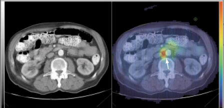
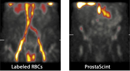
Normal distribution of labeled RBCs and a delayed ProstaScint image. There appears to be no disease.
 7
7
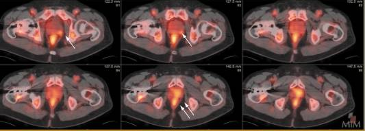
Disease is noted in the seminal vesicle (1 arrow) and in the left prostate base and midzone (2 arrows)
This patient was treated with brachytherapy seven years ago. His PSA was 1.8ng/mL. Uptake is noted in a lymph node located in the interaoctrocaval groove
http://brighamrad.harvard.edu/Cases/jpnm/hcache/1094/full.html
http://www.med.harvard.edu/JPNM/TF98_99/Oct6/Unknown.html
Return to the Beginning of the Document
Return to the Table of Content
10/14
References
1. http://www.fairview.org/healthlibrary/Article/85539 - Excellent article on Screening for prostate cancer by RM Hoffman.
2. http://leaderlinestudios.com/project/prostatecentre/ - Images and information attained at this site.
3.
Clinical experience with intensity-modulated radiation therapy (IMRT) for prostate cancer with the use of rectal balloon for prostate immobilization - http://www.sciencedirect.com/science/article/pii/S0958394702000924
4. The Cancer Center - http://drbobcole.com/prostate-cancer-treatment/prostate-brachytherapy-seed-implants/
5. Prostate Cryosurgery - http://www.prostate-cancer-miami.com/page8/prostate-cryotherapy.html
6. Cancer Research UK - http://www.cancerresearchuk.org/about-cancer/type/prostate-cancer/treatment/the-stages-of-prostate-cancer
7. ProstaScint Scan: Contemporary Use in Clinical Practice by SS Taneja - http://www.ncbi.nlm.nih.gov/pmc/articles/PMC1472938/#!po=32.1429
