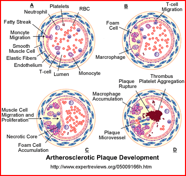
- Atherosclerosis
- The build up of fatty deposits within the lumenal wall of the coronary arteries reduces blood flow and effects myocardial function
- Fatty deposits are a result of cholesterol and fatty acids building inside the arterial wall
- These fatty streaks deposit (A) along the endothelium wall cause elongation of these flatty deposits. This can start as early as the age of 10
- Development of atherosclerotic plaque
- An intermediate lesion slowly formed (B) resulting in the invasion of macrophage foam cells. This inflammatory process extends into the lumen
- Smooth muscles then invades this sclerotic development (C) and forms a fibrous cap that contains s lipid core
- Thrombus can form (D) as the plaque ruptures causing the clot to block coronary blood flow resulting in an infarct
- Plaque/atherosclerosis
- Fibrousis is noted with B and C where muscle tissue migrates into the area of the foam cells. These macrophages reduces blood flow that cause ischemic changes in the myocardium
- Complicated lesions, such as D shows the development of a thrombus which once released may block the coronary vessel further down stream causing infarct
- Other complications may include: ulceration of the area, calcification, or hemorrhaging
- While plaque development can starts between 20 to 30 years of age, the regulating factor is the amount of cholesterol circulating in the vascular compartment. The greater its level the sooner plaque builds up occurs resulting in ischemic changes within the myocardium
- The build up of fatty deposits within the lumenal wall of the coronary arteries reduces blood flow and effects myocardial function
- Symptoms and coronary insufficiency
- Ischemic changes and its relationship to pain
- In general, as ischemia increases so does chest pain
- Related to this is the demand for oxygenated blood. At rest a highly occluded (~90%) coronary vessel may supply enough oxygen to the surrounding tissue, which means that there will be no chest pain. However, as the patient exercises, even slight, chest pain usually occurs
- Levels of chest pain
(1 through 4)
- (1) Stable angina pectoris is chest pain that occurs when the person in exercising, but goes away at rest. In this situation the occlusion is not "that bad" but it is a warning that something may be wrong!
- Interesting to note, 70 - 80% of patients with CAD are usually asymptomatic, which is partially do to normal blood flow occurs when the heart at its resting state
- (2) Unstable angina means that pain occurs at any time, rest or stress. This usually indicates a very high level of occlusion and failure of medical intervention will eventually lead to an MI
- (3) Crescendo angina is identified as continued buildup of chest pain over several weeks to a month's period of time
- (4) Acute coronary insufficiency is the final stage of unstable chest pain. This occurs all the time and at any time. If the patient's cardiac enzymes are tested or an EKG is preformed both will be abnormal
- Comment - diabetic patients may not have pain with related CAD. This condition is known as silent myocardial ischemia (How might this be a concern during an MPI procedure?)
- Why? Diabetics have nerve damage in the area of the myocardium resulting in no chest pain. This is refereed to "silent" heart disease and is defined as diabetic heart disease (DHD)
- Cardiac enzymes/cardiac biomarkers (newer terminology) - Where cardiac tissue is damaged, enzymes are released, such as troponin and creatine phosphokinase (CPK)
- The acute MI
- Symptoms - chest pain and/or pain going down the arms/elbows, diaphoretic, skin may appear pale/gray, palpitations
- Occlusion
- Once the lumen within a myocardial artery becomes completely blocked the loss of blood causes death to the myocardial tissue
- Usually irreversible damage occurs to the effected area of the myocardial wall
- Occasionally, over time, collateral circulation may re-establish cell viability to the damaged area
- Other terms associated with CAD
- Stunned myocardium - usually causes when there is an acute episode of ischemia, then the blood supply returns to normal. This results in decreased contractility to the area, however, normal cardiac function does return over time. The amount of time in which this occurs can be anywhere between days to weeks. This sudden drop or loss of blood can occur with: cardiac surgery, angioplasty, or unstable angina. How would a stunned myocardium appear in an MPI procedure?
- Hibernating myocardium - Is where the heart response to chronic reduction of blood flow, at rest, associated with reducing its cardiac contractions. It is believed to be a protective response to partial occlusion of the coronary arteries This situation requires surgical intervention. Otherwise over time an infarct will occur
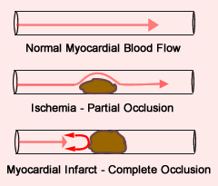
- A look at the hemodynamics
- The above slide shows the differences in blood flow specifically between normal, ischemic, and infarct
- One should appreciate that a stress test causes high flow rates and when associated with partial occlusion reduced blood flow occurs. It is this reduced flow that translates to reduced cardiac tracer uptake in the effected myocardial wall
- In addition to reduces flow there will be reduced cardiac contractility to the effected side
- Ischemic changes and its relationship to pain
- Other types of cardiac disease
(that may or may not be detected by nuclear medicine)
- Inflammatory disease
- Pericarditis
- Infection to the protective sac surrounding the heart
- Symptoms include: chest pain radiating to the back, cough, fever
- Cause may be do due to: viral, immune response (lupus or rheumatic fever), MI, trauma, other
- Enodcarditis
- Infection that involves the inner layers of the heart that usually involves the valves of the heart
- Usually causes by bacteria and may be due rheumatic fever, tooth disease, surgery, or even auto-immune
- Pericarditis
- Valvular disease
- Heart valves prevent blood from flowing in the opposite direction. When these valves become diseased, blood flows going in the opposite direction can occur. This is referred to as a "leaking" valve
- Another term is regurgitation
- Stenosis occurs when the valve fails to completely open result in some form of partial blockage
- Regurgitation and stenosis can occur at the same time
- Stenosis and regurgitation can occur with: aortic, mistral, or tricuspid valve(s)
- Pulmonary valve stenosis occurs when there is reduced blood flow to the lungs
- Nuclear medicine is generally not used to diagnose valvular disease, however, I have noticed tricuspid valve regurgitation related to a very high left ventricle ejection fraction (90%). Ultrasound is preferred when imaging the valves
- Heart valves prevent blood from flowing in the opposite direction. When these valves become diseased, blood flows going in the opposite direction can occur. This is referred to as a "leaking" valve
- Hypertension leads to hypertensive heart disease
- Can lead to heart failure where the heart becomes weaken and the walls become less elastic
- Ischemic disease can occur, which we have talked about
- Hypertrophic cardiomyopathy can occur which is where the heart walls become thicker over time, this effects blood flow through the ventricles
- Congenital heart defects occur in newborns where an actual structural defect causes alteration of in blood flow within the myocardium, which could occur with the cardiac chamber(s) or vessels
- Cardiac heart failure (CHF) may lead to heart failure
- CHF can occurs when the pumping mechanism of the heart is out of balance. Failure can be caused by
- Ischemia
- Infarct
- Wall thickening and hypertension
- Valvular disease
- Congenital defect
- In some cases edema will develop throughout the body
- The cause is related to the bodies inability to properly manage its cardiac function
- As function becomes impaired so does the lymphatic system
- This causes fluid to build up
- In the extremities, fluid builds up underneath the skin and in extreme situations pitting edema
- Pulmonary edema is noted in lungs where guild leaks into the pulmonary capillaries
- CHF can occurs when the pumping mechanism of the heart is out of balance. Failure can be caused by
- Inflammatory disease
Return to the beginning of the document
Return to the Table of Content